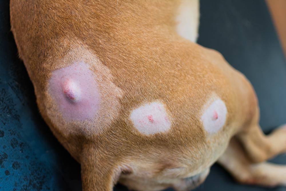
Finding a lump on your pet sets off a familiar sequence: you notice it, you poke it a few times, you worry, and then you either call the vet or talk yourself out of it. The honest reality is that no one, including veterinarians, can look at a lump from the outside and tell you with certainty what it is. A soft, moveable mass might be a benign fatty deposit. It might not be. The only way to know is to sample it, and the choice of how to do that, whether through a fine needle aspirate or a tissue biopsy, depends on where the mass is, what it looks and feels like, and what we need to know to make the next decision.
Valley Center Veterinary Clinic walks pet families through the clinical reasoning behind every recommendation, and mass evaluation is no exception. When you’re worried, we’re here to help with AAHA-accredited medical excellence paired with transparent pricing so you know you’re never left wondering. Our diagnostics include both fine needle aspiration and tissue biopsy capabilities, paired with specialist laboratory analysis, and our surgery team handles excision when the results call for it. Request an appointment or contact us to discuss a mass your pet has developed. Prompt diagnosis is critical for potentially cancerous lumps.
What Matters Most
- No one can reliably diagnose a lump by looking at or feeling it; sampling the mass with fine needle aspiration or biopsy is the only way to know whether it is benign or malignant.
- Fine needle aspiration is usually the first diagnostic step because it is quick, well-tolerated, and typically does not require sedation, while biopsy comes next when cells alone do not give a definitive answer.
- Some clinical situations including splenic masses and chronic GI disease in cats bypass aspiration entirely because reliable diagnosis requires tissue architecture that only biopsy can show.
- Benign results still warrant home monitoring with measurements and photos, since new masses can develop alongside known ones and cellular characteristics can sometimes change over time.
Why Is It Impossible to Diagnose a Lump Just by Looking at It?
The instinct to evaluate a lump visually is universal, but visual evaluation provides surprisingly little reliable information. Malignant tumors and benign masses can look and feel essentially identical. A soft, freely movable lump under the skin might be a lipoma (a harmless fatty mass) or a soft tissue sarcoma (a malignant tumor that originates in connective tissue). The only reliable way to distinguish them is sampling.
Cancer in pets is common enough that testing any new lump is routine practice rather than overreaction. Roughly one in four dogs and a substantial number of cats develop cancer at some point, and many of those cancers begin as visible or palpable masses.
The signs of cancer in pets include several presentations beyond just lumps:
- Persistent weight loss
- Decreased appetite
- Lethargy
- Changes in urination or defecation
- Sores that do not heal
- Persistent vomiting or diarrhea
- Lameness that does not resolve
For visible or palpable masses, the diagnostic question reduces to one essential step: getting cells or tissue from the mass for evaluation.
Why Do Skin Masses That Look Similar Behave So Differently?
Several common skin masses can look nearly identical on the outside but have completely different prognoses. This is one of the clearest reasons that visual evaluation alone is not sufficient. A lump that resembles a harmless fatty mass might be benign, or it might be a soft tissue sarcoma; a small bump that looks like an insect bite might be a mast cell tumor. The only way to properly diagnose a tumor is to sample.
Common Masses in Dogs Found by Pet Owners
Skin cancer in dogs takes several forms, and benign mimics of these cancers are common.
- Histiocytoma is a benign mass typically affecting young dogs (under 3 years of age in most cases). They appear suddenly as small, raised, hairless red bumps and often regress spontaneously over weeks to months, but can look just like a mast cell tumor.
- Melanocytic tumors range from completely benign to highly aggressive malignant melanomas. The location matters significantly: melanomas in the mouth or on the toes have different behavior than those on furred skin.
- Cysts in dogs include sebaceous cysts, follicular cysts, and various other types. Most are benign but they can become inflamed or infected, and get large quickly- which can also be seen in cancers.
- Mast cell tumors are one of the most common malignant skin cancers in dogs and one of the most variable in appearance. They can look like an insect bite, a small pimple, an inflamed cyst, a lipoma, or almost anything else. Boxer breeds have particularly high rates.
- Lipomas in dogs are benign fatty masses that are extremely common, particularly in older overweight dogs. The catch: soft tissue sarcomas can present with very similar physical characteristics, which is why aspirating “obvious lipomas” remains standard practice.
Common Masses Found in Cats by Pet Owners
Cats develop different skin tumor patterns than dogs, with several specific types worth knowing about:
- Feline basal cell carcinoma is one of the more common feline skin tumors, typically presenting as a slow-growing nodule on the head, neck, or trunk.
- Squamous cell carcinoma is a more aggressive cancer affecting cats, particularly white cats and those with light pigmentation in sun-exposed areas. Early lesions can look like simple sunburn or scabby spots, but by the time they become obviously concerning, they are often locally advanced.
- Fibrosarcoma in cats can develop at sites of previous injection or vaccination or arise spontaneously, and they tend to be aggressive locally with high recurrence rates after surgery.
- Feline mast cell tumors behave somewhat differently than canine mast cell tumors, occurring in cutaneous (skin) and visceral (internal) forms.
The stakes of missing a diagnosis in cats are high because feline tumors often present at advanced stages by the time you notice them.
What Is Fine Needle Aspiration and How Does It Work?
For most masses, fine needle aspiration is the appropriate first diagnostic step. It is quick, well-tolerated, and provides useful information for many situations. Most pets handle it about the same way they handle a vaccination, and results come back from the laboratory within a few business days. The procedure works best for accessible masses including skin lumps, lymph nodes, mammary masses, and some superficial organ masses by using ultrasound.
How Fine Needle Aspiration Works
Fine needle aspiration (FNA) uses a thin needle (similar in size to vaccine needles) inserted into the mass to collect cells for microscopic examination. The procedure typically takes seconds per sample. Most pets tolerate it like a vaccination without any sedation. Some pets benefit from light sedation if they are particularly anxious or if the mass is in a sensitive location.
The collected cells are placed on a glass slide, stained, and examined under a microscope. Specialist veterinary pathologists evaluate the cellular characteristics and provide a written report with their interpretation. Less accessible masses (deep within the abdomen or chest) typically require ultrasound guidance to direct the needle accurately.
What FNA Can and Cannot Diagnose
Cytology examines individual cells rather than tissue structure. It works particularly well for masses where the cellular characteristics provide clear diagnostic information. Many cancers can be definitively diagnosed by FNA alone, including mast cell tumors (which are often easily recognized by their distinctive cellular appearance) and many lymphomas.
The limitations matter: some tumor types shed cells poorly into the needle, producing non-diagnostic samples even from a malignant mass. Some cancers require evaluation of tissue architecture (how cells relate to each other in 3D space) that cytology cannot provide. And some masses are too small, too deep, or in locations where adequate sampling is difficult.
When FNA produces a non-diagnostic result or strong suspicion of malignancy persists despite benign-appearing cytology, biopsy provides the next level of evaluation.
When Is a Biopsy Needed Instead of an Aspirate?
Biopsy moves beyond cytology by sampling actual tissue rather than just cells. The information available from a biopsy goes deeper than what cytology can provide, including tumor type with greater specificity, grade indicating likely aggressiveness, invasion patterns into surrounding tissue, and surgical margin assessment when the entire mass has been removed. Biopsy comes into play when cells alone do not answer the diagnostic question, when treatment planning needs specific tumor characteristics, or when certain tumor types are simply known to require tissue evaluation.
Types of Biopsy and How They Are Performed
The main biopsy approaches:
- Punch biopsy: uses a circular cutting tool (typically 4 to 8mm) to remove a small core of tissue, useful for skin masses where representative sampling is needed but full removal is not yet planned
- Incisional biopsy: removes a piece of a larger mass for evaluation before deciding on full surgery
- Excisional biopsy: removes the entire mass, which is both diagnostic and potentially curative if the mass is benign or if margins are clean
- Needle core biopsy: uses a larger-gauge needle than FNA to collect a tissue cylinder
All forms of biopsy require sedation or anesthesia for patient comfort and accurate sampling.
What Histopathology Reveals
The veterinary pathologist evaluating a biopsy sample provides information cytology cannot:
- Tumor type with greater specificity than cytology can offer
- Tumor grade indicating how aggressively the tumor is likely to behave
- Invasion into surrounding tissues, blood vessels, or lymphatics
- Surgical margins when the mass was completely removed (whether the cancer extended to the edges of the surgical specimen)
Types of cancer in pets overviews show how histopathology categorizes the various cancers and how the resulting information guides treatment.
Histopathology is the diagnostic gold standard for cancer. When definitive diagnosis matters most, biopsy and histopathology are typically the answer.
Which Situations Require Biopsy From the Start?
Some clinical situations bypass FNA entirely and go directly to biopsy because that is the only path to reliable diagnosis. Splenic masses and chronic GI disease in cats are two of the most common scenarios where cells alone simply cannot give us a usable answer, and biopsy is built into the workup from the beginning.
Splenic Masses: Benign or Hemangiosarcoma?
Splenic masses are particularly challenging diagnostically. Imaging (ultrasound or CT) cannot reliably distinguish between benign hematomas, benign nodular hyperplasia, and malignant hemangiosarcoma in dogs.
FNA of the spleen is often unreliable. Hemangiosarcoma cells often do not aspirate well, and bleeding from the highly vascular spleen creates additional risk. Many splenic masses end up requiring splenectomy (surgical removal of the spleen) followed by histopathology of the entire removed organ to make the definitive diagnosis.
This is why decisions about splenic masses can feel uncomfortable. We are often recommending major surgery without definitively knowing whether the mass is benign or malignant. The reasoning: even benign splenic masses can rupture catastrophically, and waiting often leads to worse outcomes regardless of the underlying diagnosis.
Chronic GI Disease in Cats: IBD or Lymphoma?
This diagnostic dilemma is one of the more challenging in feline medicine. Inflammatory bowel disease in cats and feline lymphoma (specifically the small-cell GI form) can produce nearly identical clinical signs, bloodwork findings, and imaging results.
Both cause chronic vomiting, diarrhea, weight loss, and decreased appetite. Both produce thickened intestinal walls visible on ultrasound. Both can produce similar bloodwork patterns. The treatments, however, differ significantly: IBD is managed with diet and immunosuppressive medications, while lymphoma is treated with chemotherapy.
Definitive distinction requires biopsy, typically from full-thickness intestinal samples obtained surgically or via endoscopic biopsy. The biopsy diagnosis directly determines the appropriate treatment path.

Can a Blood Test Detect Cancer Before a Lump Appears?
A newer category of diagnostic testing detects cancer signals in blood before visible masses appear. Cancer screening blood tests look for genetic material from cancer cells circulating in the blood, and they can flag signals from various cancer types and provide early warning before clinical disease develops. The tests do not independently diagnose cancer; a positive result indicates that follow-up diagnostics are needed to determine where the signal is coming from.
These tests are particularly valuable for breeds at high risk of specific cancers. For example, Golden Retrievers have notably elevated rates of lymphoma and other cancers. Routine blood testing for lymphoma can pick it up months before symptoms even appear, making early diagnosis and treatment a real possibility.
The screening fits into our wellness and preventive care approach for senior pets and high-risk breeds, often integrated into annual visits. Our senior pet wellness packages include enhanced cancer screening with chest x-rays and ultrasound as part of comprehensive evaluation.
How Do I Monitor Benign Masses Over Time?
A benign result is great news, but it does not end the conversation entirely. Even confirmed benign masses warrant ongoing monitoring because new masses can develop alongside benign ones, masses that started out looking benign can change over time in some mass types, and misdiagnosis is rare but possible, particularly for sample-sensitive tumor types. A simple home monitoring routine catches changes early and gives us something to compare against if anything looks different at the next visit.
A simple monitoring routine for benign masses:
- Measure the mass in two dimensions and record the measurements
- Photograph the mass with a coin or ruler for size reference
- Date the record so changes over time are obvious
- Check monthly as part of a head-to-tail home exam
- Call us if changes occur (rapid growth, change in texture, ulceration, color change, or new pain)
Repeat aspiration is often appropriate when changes occur, even when the original diagnosis was benign. Some masses that are benign will continue to grow. Lipomas are the prime example of this, and can grow to be so large they actually impede your pet’s normal movement and become extremely uncomfortable, warranting removal even though they are technically non-cancerous.
What Happens After Diagnostic Results Come Back?
Three result categories with different next steps:
- Benign results establish a monitoring plan. The mass is documented, monitored at home, and rechecked at routine visits. No treatment is typically needed unless the location or size becomes problematic.
- Malignant results open the treatment conversation. Depending on the specific cancer, the next steps might involve surgical removal with appropriate margins, additional diagnostics for staging (looking for spread to other locations), oncology consultation for treatment planning, or palliative care decisions. Different cancers have very different treatment approaches and prognoses, so the conversation gets specific quickly.
- Inconclusive results guide further diagnostics. If cytology was non-diagnostic, biopsy may be the next step. If a mass appears different than the cytology suggests, repeat sampling or alternative approaches may help.
For the cases requiring difficult decisions, our end-of-life services provide compassionate support when treatment is not the right path.
Frequently Asked Questions About Cytology and Biopsy
Does my pet need sedation for an FNA?
Usually no. Most pets tolerate FNA like a vaccine. Some benefit from light sedation if anxious. Biopsy, on the other hand, requires sedation or anesthesia.
How long does it take to get results?
FNA results typically come back within 2 to 4 business days. Biopsy results often take 5 to 10 business days because of the additional processing required.
Can a benign mass become malignant?
Sometimes, though most do not. Some specific mass types have small potential for malignant transformation, which is why monitoring matters.
What is the cost difference between FNA and biopsy?
FNA is significantly less expensive because it does not require sedation, surgical prep, or extensive lab processing. The cost difference is one reason FNA is typically the first step.
Finding Answers Sooner for Your Pet
Earlier testing means more options and better outcomes. The lump that turns out to be benign produces relief; the one that turns out to be malignant typically has more treatment options when caught early. Either outcome is better than waiting in uncertainty.
If you have found a new lump on your pet, request an appointment at Valley Center Veterinary Clinic. Our team will work through evaluation systematically and develop a clear next step regardless of what the testing reveals.


Leave A Comment